Research
Interests
Last updated: 8/16/2013
- Characterizing/developing molecular
interaction using a QCM-D (Quartz crystal microbalance with
dissipation) sensor
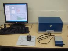
A sensing unit of QCM-D sensor is a thin quartz
crystal (diameter 2.5 cm, thickness 1mm) with thin gold electrodes coated in
both sides. The crystal is piezoelectric
resonators where the resonant frequency varies linearly with the mass of
adsorbed layers on the surface when it is in contact with air. After it was shown that the QCM might be
used in the liquid phase, the number of applications for the QCM has increased
dramatically. In liquid, an adsorbed film may consist of a considerably high
amount of water, which is sensed as a mass uptake. By measuring several frequencies and the
dissipation it becomes possible to determine whether the adsorbed film is rigid
or water-rich. The QCM-D sensor monitors
dissipation, allowing biomolecular detection in liquid. Read more in www.qsense.com
- SAM (Self Assembled Monolayer) on the
gold substrate
It
is critical to develop a reliable deposition method that can ensure sufficient
amount of protein/peptides can be deposited onto the QCM surface in a controllable
manner. Several strategies are known to
effectively deposit proteins onto the sensor surface. They include direct adsorption onto citrate
coated surface, amine or thiol coupling onto self assembled monolayers (SAM) or via linkage of
streptavidin – biotin.
- DNA sensing using the QCM-D and EIS
Detection
of specific DNA/RNA sequences is important in numerous applications including
clinical diagnosis such as genetic disorders and pathogen detection. The detection of mismatched base pairs in DNA
plays a crucial role in the diagnosis of genetic-related diseases and
conditions, especially for early stage treatment. The Quartz Crystal
Microbalance with Dissipation (QCM-D) sensor combined with electrochemical
impedance spectroscopy (EIS) are used to monitor DNA
hybridization and mismatched base pair detection. The QCM-D technique measures the adsorbed
mass change while EIS gives additional electrochemical signal change.
3.
Simultaneous QCM-D and Extended gate Field Effect
Transistor (EGFET)
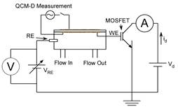 A
merged setup of quartz crystal microbalance
with dissipation (QCM-D) sensor and extended gate field effect transistor
(EGFET) was used to monitor biomolecular interactions. The biosensor simultaneously
provides multiple information on
electrochemical, thickness/flexibility, and structural changes which accompany
bio-molecular interactions. The
sensing environment is separated from FET by wiring the gate
to the external working electrode- the gold electrode of the QCM-D device- and
thus reduce solution interference.
The setup utilizes a commercial,
affordable MOSFET, hard wired to the QCM-D setup without having to undergo
complicated nanofabrication procedures.
In this paper, characterization of I-V of the novel EGFET setup, monitoring
of avidin-biotin, and binding between calmodulin and its
binding moieties are presented.
Combined electrochemical and thickness data clearly demonstrated data
acquisition on electrochemical change, adlayer
thickness change and structural change upon binding. .
A
merged setup of quartz crystal microbalance
with dissipation (QCM-D) sensor and extended gate field effect transistor
(EGFET) was used to monitor biomolecular interactions. The biosensor simultaneously
provides multiple information on
electrochemical, thickness/flexibility, and structural changes which accompany
bio-molecular interactions. The
sensing environment is separated from FET by wiring the gate
to the external working electrode- the gold electrode of the QCM-D device- and
thus reduce solution interference.
The setup utilizes a commercial,
affordable MOSFET, hard wired to the QCM-D setup without having to undergo
complicated nanofabrication procedures.
In this paper, characterization of I-V of the novel EGFET setup, monitoring
of avidin-biotin, and binding between calmodulin and its
binding moieties are presented.
Combined electrochemical and thickness data clearly demonstrated data
acquisition on electrochemical change, adlayer
thickness change and structural change upon binding. .
- Olfactory signal transduction
mechanism
My
post-doc research topic was elucidating a role of olfactory marker protein
(OMP) in the olfactory signal transduction cascade. Using the confocal imaging of intracellular
Ca2+ in the living olfactory tissue, the intake and extrusion of Ca2+
was studied in normal and OMP-KO mice.
Study of enigmatic OMP is still on-going quest.
·
Modeling
and Simulation Study
Simultaneous fluid, diffusion-convection, and mass adsorption model in the biosensor was developed and studied using the COMSOL Multiphysics. Effects of feed concentration, flow rates, and binding rate constants on the sensor gram are often simulated. The study can be used to optimize the sensing conditions and guide determination of the affinity upon biomolecular interaction. The study can be used to optimize the sensing conditions and guide determination of the affinity upon biomolecular interaction. See details in http://www.comsol.com/papers/6561/
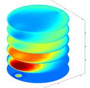
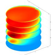
Transient concentration profile of the species B (left) and bound complex (right) at the upper
surface for time points of 1, 15, 30, 45, 60, 75 seconds.