|
1
|
- Fundamental Concepts in Immunology
- Program for Clinical Laboratory Science
- Unit - 07
- Abnormal Immune Responses
|
|
2
|
- Reading assignment:
- Pages 148 - 171 of textbook
- Learning objectives:
- Those listed on page 149 of textbook
- Key terms:
- Those listed on pages 149 & 150 of textbook
|
|
3
|
- Hypersensitivity Disorders
- p
- Autoimmune Disorders
- p
- Immunodeficiency Disorders
- p
- Hypergammaglobulinemia Disorders
|
|
4
|
- Hypersensitivity Disorders
|
|
5
|
- Definition:
- a normal but exaggerated or uncontrolled immune response to a persistent
antigen and results in inflammation and tissue damage.
- Four types of hypersensitivity disorders:
- Type I
- Type II
- Type III
- Type IV
- Immediate or allergic hypersensitivity
- Antibody-Dependent Cytotoxic hypersensitivity
- Immune Complex-Mediated hypersensitivity
- T-cell mediated hypersensitivity
|
|
6
|
- Description:
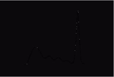
- an immune response (called an allergic reaction or allergy) that is
mediated by IgE and occurs immediately after subsequent contact with
same antigen (called allergen).
- Mechanism of tissue damage:
- Uduring 1st contact with allergen IgE production is initiated by TH2
response to allergen.
- UIgE produced bind to mast cells via Fc receptors and mast cells are
said to be sensitized.
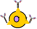
- mast cell
- Fc receptors
- IgE
- Sensitized mast cell
|
|
7
|
- Mechanism of tissue damage: - cont=d
- Uin subsequent allergen exposure the allergen binds with IgE on
sensitized mast cells
- allergen must bind to 2 or more IgE molecules before mast cell will
secrete mediators:
- eosinophil chemotactic factor
- histamine
|
|
8
|
- ]localized reaction:
- -a cutaneous reaction with:
- La Awheal and flair@ reaction
- Llocal edema
- Litching at site of allergen introduction
- ]generalized reaction:
- -also known as Aanaphylaxis@
- -vasodilation
- -bronchioconstriction
- -edema
- Miscellaneous information:
- Uthe allergic reaction may be induced by C3a and C5a
- Uthe level of free IgE can be measured by RAST to determine allergen
responsible for reaction
|
|
9
|
- Description:
- an immune response that occurs when antibody (IgM or IgG) binds with
cell surface antigen and activate complement which lyse cells and the C3a
and C5a attract macrophages, neutrophils, and basophils into area or NK
cells lyse antibody-coated cells.
- Mechanism of tissue damage:
- Uantigen causes antibody to be produced
- Uantibody produced attached to antigen on target cell
- UAb-Ag complex may activate complement which lysis target cell via MAC
attack
- and/or
- UAb-Ag complex activates NK cell or TCytotoxic cells which
lysis target cell
|
|
10
|
|
|
11
|
- Antibodies are formed that bind to specific target cell antigens and the
antibody-coated target cells are destroyed by cytotoxic substances being
released from effector cells which bind via Fc receptors to the antibody=s
Fc portion.
|
|
12
|
- Blood Transfusion Reactions
- Š
- Hemolytic Disease of Newborn (HDN)
- Š
- Goodpasture=s Syndrome (Glomerulonephritis)
|
|
13
|
- Uthe reaction occurs when:
- ]incompatible antigens on donor red blood cells are infused into
recipient
- ]recipient=s immune system has produced antibodies against antigens from
a previous exposure
- Uthe reaction=s features :
- ]previously produced antibodies form Ab/Ag complexes on RBC=s surface
- ]these complexes activate complement via classical pathway
- ]RBC=s destroyed via MAC intravascularly
|
|
14
|
- Uthe reaction occurs when:
- ]IgG antibodies from mother cross placental barrier and attack red cells of fetus
- ]mother must have been exposed to red cell antigens prior to pregnancy
(previous pregnancy or transfusion)
- ]the incompatible antigens on fetal red cells are inherited from father
- Uthe reaction=s features :
- ]Ab/Ag complexes are formed on fetal red cells
- ]fetal red cells are destroyed
|
|
15
|
- Uthe reaction occurs when:
- ]antigen/antibody complexes form along basement membranes of glomeruli
and alveoli
- ]complement is activated via classical pathway by complexes and MAC is
responsible for tissue damage
- ]neutrophils accumulate at site of injury and release enzymes adding to
injury
- Uthe reaction=s features :
- ]autoantibodies formed against collagen of basement membranes
- ]lesions form in both lungs and kidneys
|
|
16
|
- Serum Sickness
- Š
- Systemic Lupus Erythematosus
- (SLE)
|
|
17
|
- Antigen/antibody complexes formed are not effectively removed by
phagocytes and are deposited in various tissues and organs.
- Tissue damage occurs at the site of immune complex deposition.
|
|
18
|
- Autoimmune disorders
- Persistent infection
- Repeated inhalation of antigens
- rhematoid arthritis
- viral hepatitis
- moldy hay
- Farmer=s lung
|
|
19
|
- Uincreased vascular permeability
- histamine
- Uhigh blood pressure and turbulence
- in glomerular capillaries
- Usize of antigen
- larger antigens form larger complexes
- Uaffinity of antigen for tissue
- basement membranes & synovial linings
- Uclass of antibody involved
- IgG and anti-IgG
|
|
20
|
- Ucomplement is activated releasing anaphylotoxins (C3a & C5a)
- Uanaphylotoxins stimulate mast cells and basophils to release
chemotactic factors & amines (histamine)
- Ueffector cells are called into area which release their lysosomal
enzymes
- Ua local inflammatory reaction occurs
- Ubegins when Ag/Ab immune complexes are deposited onto tissue
|
|
21
|
- Udecreased serum complement levels
- Udetection of immune complexes
|
|
22
|
- Uself-limiting systemic immune complex disease affecting:
- lymph nodes
- joints
- UCaused by passive immunization
- administration of pre-formed antibodies (gammaglobulin shots) formed in
horses
- UMechanism:
- proteins from horse serum causes host to produce antibodies that
complexed with horse proteins forming immune complexes
|
|
23
|
- Uan autoimmune disease characterized by:
- presence of autoantibodies
- immune complexes
- UImmune complexes are deposited within kidney glomeruli
- results in inflammation of blood vessels within the glomeruli
|
|
24
|
- Examples:
- Allergic contact dermatitis
- Š
- Tuberculin & Other skin tests
- Other Name:
- T cell-mediated hypersensitivity
|
|
25
|
- Mediated by T lymphocytes which had previous exposure with a stimulating
antigen and release cytokines which induce an inflammatory reaction,
activate and attract macrophages into area.
- Inflammation and swelling of infected area results.
|
|
26
|
- Sensitization phase
- Š
- Effector phase
|
|
27
|
- U lasts for 1 - 2 weeks following initial contact with antigen
- UT-helper cells are activated and proliferate when antigen is presented
by APC=s in conjunction with MHC -II
- UT-helper cells are activated and proliferate when antigen is presented
by APC=s in conjunction with MHC -II
- UT-cells that are activated are designated as TDTH
|
|
28
|
- U does not show clinical symptoms until 24 hours following subsequent
contact with antigen
- Upeak response occurs between 48 - 72 hours
- UT-DTH cells secrete a
variety of cytokines that are responsible for :
- erecruitment & activation of macrophages
- erecruitment & activation of other non- specific inflammatory cells
- UGenerally the antigen is cleared with little tissue damage
- UIf the antigen is not cleared easily, a granuloma may develop
|
|
29
|
- UT-DTH cells secrete a
variety of cytokines that include the following:
- eIL-3
- eGM-CSF
- eINF-(
- eTNF-$
- eMIF
- chemotactic for macrophages
- hematopoietic stimulus for monocytes & neutrophils
- affects permeability of endothelial cells and activates macrophages
- migration-inhibition factor inhibits macrophages from leaving area of
DTH reaction
|
|
30
|
- Ualso called allergic contact dermatitis
- Ucharacterized by:
- eskin eruptions
- elesions of skin
- eblistering
- escaling
- eedema
- Udiagnosis confirmed by Apatch testing@
- Umay persist for years or even a lifetime
|
|
31
|
- Usome causative agents include:
- epoison ivy or poison oak
- ehair dyes
- eformaldehyde
- enickel
- eturpentine
- evarious cosmetics
|
|
32
|
- Uperformed by injecting of test antigens intradermally
- Umost common allergins injected include:
- ePPD - purified protein derivative from M. tuberculosis is used
- eCandida albicans
- Utests read between 48 - 72 hours after injection
- Udevelopment of a red, slightly swollen firm lesion indicates a positive
reaction
|
|
33
|
|
|
34
|
- When the immune regulatory mechanism becomes responsive to Aself-antigens@
giving rise to an uncontrolled and exaggerated immune response
- The autoimmune response is an antigen- specific immune response that may
be either cell-mediated or humoral
|
|
35
|
- Uinvolves the T lymphocyte populations
- UPre-T lymphocytes with a potential to react with self-antigens are
eliminated in the thymus gland
- Examples of Self-antigens involved in autoimmune disorders include:
- Ucell-surface receptors
- UErythrocyte surface proteins
- UBasement membranes of glomeruli
- UHormone receptors
- UNucleoproteins
|
|
36
|
- Alterations in lymphocytes
- |
- Local tissue alterations
- |
- Genetic factors
- |
- Environmental factors
|
|
37
|
- Uinvolves the T and/or B lymphocyte populations
- Umay invlove the following alterations or changes:
- eabnormal selection of lymphocytes
- eCross-reactions with Ashared antigens@
- eIncreased production of cytokines
- ePolyclonal stimulation of lymphocytes
- Local tissue alterations
- Uinvolves the T and/or B lymphocyte populations
- Umay invlove the following alterations or changes:
- eRelease of previously Ahidden@ self-antigens
- eAlterations in structure of self-antigens
|
|
38
|
- Uno direct evidence in humans
- Uthose that inherit certain MHC alleles have higher probability of
developing diseases
- HLA allele
- Disease
- Relative risk
- NOTE: Relative risk is the Atimes
higher@ the risk compared to normal population
- DR4
- B27
- DR2
- DR3
- DR3/DQW8
- pemphigus vulgaris -
- blistering skin disorder
- 5
- Ankylosing spondylitis
- 90
- Goodpasture=s syndrome
- 16
- Myasthenia gravis
- 10
- Diabetes mellitus
- 100
|
|
39
|
- Usome factors that may cause alterations in self- antigen
- eDrugs
- eToxins
- ËPenicillin - autoimmune Hemolytic anemia
- ËProcainamide - SLE
- ËMercuric chloride - antinuclear antibodies
- ËPolyvinyl chloride - scleroderma
- ËSilicon - scleroderma & Rheumatoid arthritis
- eUltraviolet radiation
- Ëcorrelated to increased risk for skin cancers but not with autoimmune
disorders
|
|
40
|
- Organ-Specific
- Š
- Organ-Non-Specific
|
|
41
|
- The immune response is directed primarily against antigens in or on a
specific organ
- Organ involved
- Antigen targeted
- Disease
- Clinical features
- Adrenal gland
- Adrenal cortex
- Addison=s
- Hypoadrenalism
- Thyroid gland
- TSH receptors
- Grave=s
- Hyperthyroidism
- Thyroid cells
- Hashimoto=s
- Hypothyroidism
- Skeletal or heart Muscle
- Acetylcholine receptors
- Myasthenia gravis
- Muscle weakness
- Kidney
- Glomerular basement membrane
- Goodpasture=s
- Glomerulonephritis
- Pancreas
- Islets of Langerhan
- Diabetes mellitus type I
- Insulin-dependent hyperglycemia
- CNS
- myelin
- Multiple sclerosis (MS)
- neurologic dysfunction
|
|
42
|
- The immune response is directed against specific antigens but the immune
complexes involve many other organs or tissues of the body
- Examples of these types of diseases include:
- Systemic Lupus Erythematosus (SLE)
- Š
- Rheumatoid Arthritis (RA)
|
|
43
|
- Mechanism responsible for tissue injury include:
- UAntibody-mediated cytotoxicity
- eIgG or IgM type antibodies
- eAttach to antigen and causes cytolysis as in type II hypersensitivity
reactions
- UImmune complex deposition
- eAntibody-Antigen complex forms
- eNormally removed by phagocytes
- eIf not removed they are deposited on various tissues as in Type III
hypersensitivity reactions
- UCell-mediated cytotoxicity
- eWhen T cells fail to recognize self-antigens they can begin destroying
host cells via type IV hypersensitivity reactions
|
|
44
|
|
|
45
|
- A defect or deficiency in any one or more of the main components of the
immune system which results in an abnormally low immune response.
- The components of the immune system include:
- B lymphocytes
- Š
- T lymphocytes
- Š
- Phagocytes
- Š
- Complement
|
|
46
|
- Primary immunodeficiencies
- Šalso called congenital or hereditary immunodeficiencies
- Šimmunodeficiencies that are the result of genetic defects
- Šbecome evident at birth or early childhood
- Secondary immunodeficiencies
- Šalso called acquired immunodeficiencies
- Šimmunodeficiencies that develop subsequent to infection, cancer, or
therapy
- Šbecome evident later in life
|
|
47
|
- Name of Disorder
- Origin of Defect
- Specific Defect
- Bruton=s agammaglobulinemia
- mutated gene on X chromosome
- blocked B cell maturation
- IgG subclass deficiency
- defect in Ig isotype switching
- blocked B cell differentiation
- Common variable immunodeficiency
- intrinsic B cell defect
- B cell differentiation to plasma cells abnormal
- DiGeorge=s syndrome
- abnormal thymus gland
- defect in T cell maturation
- SCID (autosomal rec.)
- PNP enzyme deficiency
- abnormal T & B cell development
- SCID (X-linked)
- IL-2 receptor gene mutation
- abnormal T cell growth & development
- Chronic granulomatous disease
- intracellular H2O2 not produced
- defective phagocytosis
|
|
48
|
- Name of Disorder
- Origin of Defect
- Specific Defect
- Acquired Immunodeficiency Syndrome (AIDS)
- HIV-induced loss of CD4+ cells
- lysis of CD4+ cells by viral budding
- Acquired Immunodeficiencies - secondary to Malignancy or Rx
- Hodgkin=s disease
- T cell abnormality
- impaired T cell function
- Immunosuppressive drug therapy
- destruction of lymphs, PMN, & monocytes
- cytotoxic effects
- Name of Disorder
- Origin of Defect
- Specific Defect
|
|
49
|
- Characteristics of deficiency:
- Šincreased susceptibility to infection by pyogenic and intracellular
bacteria
- Šincreased tendancy to develop immune complex diseases
- Examples of some component deficiencies:
- ŠC3 - lack of opsonization results in decreased phagocytic function
- Šfactor H
- may be fatal due to pyogenic bacteria
- ŠC5 - results in lack of MAC - susceptible to intracellar infections by Neisseria
species
- ŠC1, C4, or C2 - lack of clearing of immune complexes
- Šfactor I
|
|
50
|
|
|
51
|
- An increased proliferation of antibody- producing plasma cells that
leads to increased immunoglobulin production
- Types of gammopathies:
- Monoclonal gammopathies
- Š
- Polyclonal gammopathies
|
|
52
|
- Ualso called plasma cell dyscrasias
- Ucharacterized by an uncontrolled proliferation of a single clone of
plasma cells at the expense of other clones
- Uin Multiple Myeloma the monoclonal antibodies produced in 52% of
patients is of the IgG class
- Uin Waldenstrom=s macroglobulinemia the antibodies produced are of the
IgM class
|
|
53
|
- Ucharacterized by an uncontrolled proliferation of several clones of
plasma cells which results in the production of more than one class of
immunoglobulin being produced
- UExamples of diseases associated with polyclonal gammopathies include:
- Šacute infections
- Šchronic infections
- Šrheumatoid arthritis
- Šliver disease
|
|
54
|
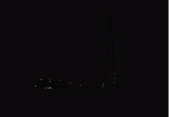
- Uprotein electrophoresis is most common method
- Normal
- Electrophoretic mobility
- -
- +
- (
- $
- "2
- "1
- globulins
- albumin
- IgG
- IgA
- IgD
- IgM
|
|
55
|
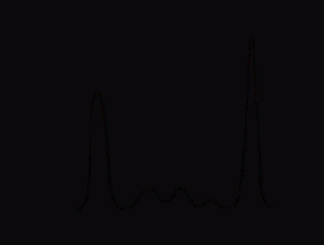
- Electrophoretic mobility
- -
- +
- Concentration º
- (
- $
- "2
- "1
- albumin
|
|
56
|
- Electrophoretic mobility
- -
- +
- Concentration º
- (
- $
- "2
- "1
- albumin
|
|
57
|
- Press the ESC key to end program
|

 Notes
Notes