|
1
|
- Diagnostic Laboratory Immunology
- Program for Clinical Laboratory Science
- Unit - 11
- Precipitation Techniques
|
|
2
|
- Reading assignment:
- Pages 228 - 252 of textbook
- Learning objectives:
- Those listed on page 229 of textbook
- Key terms:
- Those listed on pages 229 - 230 of textbook
|
|
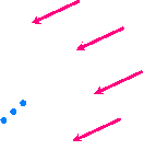
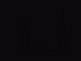
3
|
- Definition:
- Immunoprecipitation is the formation of insoluble Ag-Ab complexes called
Aprecipitins@, when soluble antigen is mixed with it=s specific antibody.
- Factors that must be considered when performing immunoprecipitation
techniques:
- Urelative concentrations of antigen and antibodies
- Uhydrogen ion concentration (pH)
- Uionic strength of solution or reaction media
- Uantibody affinity and avidity
|
|
4
|
- Immunodiffusion techniques:
- Immunoelectrophoresis techniques:
- Light Scattering Immunoassay techniques:
- Usingle immunodiffusion (Oudin)
- Udouble immunodiffusion (Ouchterlony)
- Uradial immunodiffusion
- Uimmunoelectrophoresis (IEP)
- Uimmunofixation electrophoresis (IFE)
- Uelectroimmunodiffusion
- \countercurrent electrophoresis
- \rocket electrophoresis
- Uisoelectric focusing
- Uimmunoturbidimetric methods
- Uimmunonephelometric methods
|
|
5
|
- Definition:
- Immunodiffusion is the movement of antibody molecules and antigen
molecules within a support medium
- When the two reactants meet insoluble Ag-Ab complexes are formed
(precipitins) which are visible and fixed at site of precipitation
- Antigen
- Antibody
- precipitin
|
|
6
|
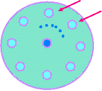
- Also called Ouchterlony procedure
- UPattern of Identity
- Continuous precipitin lines that merge and form an arc indicate
antibodies are precipitating identical epitopes
- antibody
- antigen 1
- antigen 2
- precipitin line
|
|
7
|
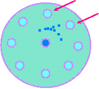
- UPattern of Partial Identity
- fusion of precipitin lines indicate antibodies are precipitating identical
epitopes common to both antigens while the spur indicates an additional
epitope not common to both
- antibody
- antigen 1
- antigen 2
- precipitin line
- precipitin spur
|
|
8
|
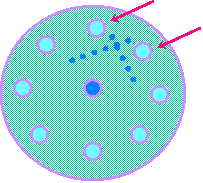
- UPattern of Non-Identity
- fusion of precipitin lines indicate antibodies are precipitating non-identical
epitopes of antigens
- antibody
- antigen 1
- antigen 2
- precipitin line
- precipitin line
|
|
9
|
- Immunodiffusion Techniques
- Double Immunodiffusion Technique (Ouchterlony):
- UPractical application:
- The Alberta Health Dept. would
like to confirm that the chicken chop suey from Wings cafJ is really
chicken.
- chicken chop suey
- anti-rabbit
- anti-cat
- anti-dog
- anti-chicken
- precipitin line
- precipitin line
|
|
10
|
- eMixed Connective Tissue Disease
- an autoimmune disorder in which antibodies react with a variety of
tissues and looks like:
- SLE - nuclear proteins
- scleroderma - blood vessels & connective
- polymyositis - muscle
- eSystemic Lupus Erythematosus
- an autoimmune disorder in which antibodies react with a variety of
tissues
|
|
11
|
- eAlteration of precipitin lines by:
- 1.excessive condensation in wells
- 2.improper filling of wells
- 3.incubation in tilted position
- efalse results due to misidentification of wells
- efalse negative results due to under incubation
- efalse results due to incorrect interpretation of pattern
- einvalid controls or standards used
|
|
12
|
- Also called Oudin procedure:
- One of the reactants is incorporated into the medium and is in fixed
position and concentration while the other reactant diffuses through the
medium from wells cut into medium.
- agar with Ab=s to Ag being tested
- Standards of differing concentrations added to wells a - d
- a
- b
- c
- d
- precipitin rings
- the larger the ring the greater the conc. of Std.
|
|
13
|
- A standard curve can then be developed based on concentration vs ring
diameter.
- 1
- 2
- 3
- 4
- 0
- b
- c
- d
- a
- Diameter (mm)
- Concentration (mg/dL)
- X
- X
- X
- plot ring size for each concentration
- join dots to form std curve
|
|
14
|
- Patient samples now can be run using same agar media
- Patients a through d added to individual wells
- a
- b
- c
- d
- The size of each ring is then plotted on std curve to determine
concentration of each patient=s antigen
|
|
15
|
- A standard curve can then be used to convert ring size to concentration
- 1
- 2
- 3
- 4
- 0
- 1
- 2
- 3
- 0
- X
- X
- X
- Diameter (mm)
- Concentration (mg/dL)
- let=s say patient c has a ring size of 3 mm
- patient c has a conc. of 3 mg/dL
- let=s say patient a=s ring size = 2.5 mm
- = 2.3 mg/dL
- let=s say patient b=s ring size = 2 mm
- = 2.0 mg/dL
- let=s say patient d=s ring size = .5 mm
- = 0.5 mg/dL
|
|
16
|
- eEnd-Point Method (Mancini):
- eKinetic Method (Fahey & McKelvey)
- 1.antigen allowed to diffuse until completion - about 24 hours
- 2.the ring diameter is directly proportional to concentration of antigen
- 3.standard curve used to convert ring diameter to concentration
- 1.antigen allowed to diffuse for limited time - about 18 hours
- 2.the ring diameter is proportional to the log of antigen concentration
- equivalence point is reached
- equivalence point is not reached
|
|
17
|
- Clinical Applications:
- Identification and quantification of serum proteins:
- -immunoglobulins (IgG, IgM, IgE, IgD, IgA)
- -"1-antitrypsin
- -transferrin
- -complement components (C3, C5, etc.)
- Technical errors:
- incorrect filling of wells
- specimen contamination
- wrong media or outdated media
- incorrect construction of standard curve
- incorrect incubation time or plate position
- incorrect measuring of ring size or calculations
|
|
18
|
- General concept:
- By applying voltage through a support medium it is possible to separate
complex protein mixtures
- Types of support media used in clinical applications:
- Upaper
- Uagarose gel
- Ucellulose acetate
- Factors influencing protein molecule migration:
- Usurface charge on molecule
- Umolecular weight of molecule
- Usize of molecule
- Umedium characteristics
- Ubuffer characteristics (pH)
- Utemperature
|
|
19
|
- Serum, urine, or CSF is applied to support medium and voltage is applied
- point of application
- anode (+)
- cathode (-)
- a stain is added that reacts with separated proteins to form a visible
band of color
- the strip is then placed in a densitometer which converts each band into
a peak based on intensity of color and width of band
|
|
20
|
- anode (+)
- cathode (-)
- (
- $
- "2
- "1
- albumin
|
|
21
|
- anode (+)
- cathode (-)
- (
- $
- "2
- "1
- albumin
- Waldenstrom=s macroglobulinemia
- Multiple myeloma
|
|
22
|
- anode (+)
- cathode (-)
- (
- $
- "2
- "1
- albumin
- chronic infection
- chronic inflammation
- chronic liver disease
- if on CSF it may indicate oligoclonal bands as seen in multiple
sclerosis
|
|
23
|
- UArtifacts from hemolyzed specimens causes increases in:
- -"2-globulin peak
- -$-globulin peak
- Upoor or excessive migration due to:
- -wrong amount of time allowed for test to run
- -wrong amount of voltage applied
- -wrong pH due to bad buffer
- Uexcessive sample applied will cause:
- -increased peaks
- -cross contamination of adjacent peaks
- Uincreased peak in $-globin area may be due to:
- -presence of fibrinogen may be mistaken for increased IgA levels
|
|
24
|
- Waldenstrom=s Macroglobulinemia
- On pages 239 - 241AOne Step Further@ presents a more in- depth
discussion of the Waldenstrom=s Macroglobulinemia.
- This presentation is contained on a separate slide presentation called A
One Step Further #10"
- Cytokines
- The student may call up the slide program OSF-10 later or click on the
arrow below to view slides now.
|
|
25
|
- Multiple Sclerosis
- On pages 243 - 244AOne Step Further@ presents a more in- depth
discussion of Multiple Sclerosis.
- This presentation is contained on a separate slide presentation called A
One Step Further #11"
- The student may call up the slide program OSF-11 later or click on the
arrow below to view slides now.
- Cytokines
|
|
26
|
- Utypically performed as confirmation when a monoclonal component is
detected on protein electrophoresis.
- Uallows for characterization of monoclonal proteins by identifying:
- -specific heavy-chain classes
- -specific light-chain components
- Ua two-step process that involves:
- -electrophoretic separation of proteins in serum or urine
- -immunodiffusion/precipitation of separated proteins with antiserum
|
|
27
|
- Userum or urine placed in antigen wells in a test gel.
- Utheir proteins are then seperated using protein electrophoretic
technique.
- Uantisera (monospecific reagent antibodies) then placed in trough in
gel.
- Uantisera and protein fraction diffuse towards each other and form
precipitin lines when equivalence point is reached.
|
|
28
|
- gel plate
- Ag well
- antisera trough
- serum added
- anode (+)
- cathode (-)
- separated proteins
- separated proteins
|
|
29
|
- monospecific antisera added
- -incubation in humid chamber for specific time
- precipitin lines form
- -position of precipitin lines compared to control for identification
- -increased in size of precipitin line indicates abnormal production of
antibodies
- -decreased in size of precipitin line indicates immunodeficiency
|
|
30
|
- IFE = Immunofixation Electrophoresis
- UUsed as an alternative to immunoelectrophoresis
- UA two step procedure as is IEP.
- UDiffers from IEP in:
- \the antisera is directly overlayed onto gel instead of
- being put in a trough
- \more sensitive
- \shorter incubation time (2 hours compared to overnight)
- \more expensive and labor-intensive
|
|
31
|
- EID = Electroimmunodiffusion
- UCountercurrent electrophoresis
- \similar to that of double immunodiffusion with the addition of an
electric current to aid in seperation of proteins
- EIA = Electroimmunoassay (Rocket electrophoresis)
- \depends on the creation of different charges of antibody and antigen at
selected pH
- \as antigen moves through gel several precipitin bands appear forming Arockets@.
|
|
32
|
- General concept:
- UBy creating a pH gradient within the medium immunoglobulin molecules (amphoteric)
will move in an electrophoretic system to a point where their
isoelectric point is equal to the pH of medium
- amphoteric molecule = molecules that can act as either a base or an acid
depending on pH of medium in which they are suspended.
- Clinical application:
- U identification of oligoclonal bands as in:
- \multiple sclerosis
- \viral encephalitis
- \cerebral infarction
- \AIDS
|
|
33
|
- General concept:
- Uwhen ag-ab complexes are formed in a solution they form
immunoprecipitates which have the ability to scatter a light beam that
passes through the solution.
- Turbidimetric application:
- U detectors set at 180o angle measures reduction in light
- Nephelometric application:
- U detectors set at 90o angle measures increases in light
- in both techniques standard curves are constructed to convert absorbance
to concentration
- in turbidimetry the absorbance is directly proportionate to
concentration so std curve is linear and fewer standards needed
- in nephelimetry the standard curve is non-linear and requires
calibrators
|
|
34
|
- Press the ESC key to end program
|
|
35
|
- Waldenstrom=s Macroglobulinemia
- OSF - 10
- Pages 239 & 241
- 7click arrow to return to main program
|
|
36
|
- Classification:
- ea monoclonal gammopathy with malignant lymphoproliferation
- Monoclonal gammopathies:
- euncontrolled proliferation of a single clone or line of plasma cells
- ealso called Aplasma cell dyscrasia@
- eplasma cell clone produces a homogenous monoclonal protein (M-protein)
- ein the case of Waldenstrom=s the clone produces a homogenous monoclonal
protein of IgM class immunoglobulins
|
|
37
|
- Mechanism of Disease:
- ecause unknown
- egenetic predisposition may play a role
- emainly seen in older individuals
- epresents with the following features:
- Žmalignant proliferation of B cell line
- Žincreased plasma cell population
- Žhigh levels of IgM produced
- Žweakness, fatigue, nose & gum bleeds, and GI bleeds leads to anemia
- Žincreased infections
- Žweight loss
- Žincreased blood viscosity
- Žmay develop neurological and cardiac problems
|
|
38
|
- Laboratory Findings:
- epresence of M-protein on serum electrophoresis
- eserum & urine characterization of M-protein using:
- Žimmunofixation test (IFE) - one of choice
- Žimmunoelectrophoresis test (IEP)
- equantification of serum IgM levels using:
- Žnephelometry procedures
- eM-protein of Waldenstrom=s may be:
- Žin serum:
- wIgM heavy chains
- weither kappa or lambda light chains
- Žin urine:
- weither kappa or lambda light chains
- wfree light chains are called ABence-Jones
proteins@
|
|
39
|
- Testing Protocol:
- ŽScreening tests
- Serum Protein Electrophoresis (SPEP)
- monoclonal bands present?
- NO
- no monoclonal gammopathy present
- YES
- Perform confirmatory testing
- to next slide
|
|
40
|
- Testing Protocol:
- ŽConfirmatory tests
- Urine and serum specimens
- CHARACTERIZATION OF MONOCLONAL BAND
- QUANTIFICATION OF IMMUNOGLOBULINS
- Serum IFE
- ID type of heavy chains and light chains
- Urine IFE
- Confirm Bence-Jones protein
- Serum Neph
- determine conc. of Ig=s
- Urine Neph
- determine conc. of light chains
|
|
41
|
- 7click arrow to return to main program
|
|
42
|
- Multiple Sclerosis
- OSF - 11
- Pages 243 & 244
- 7click arrow to return to main program
|
|
43
|
- Mechanism of Disease:
- ecause is unknown
- ebelief is that genetic and environmental factors play a role
- ŽGenetic factors:
- these two HLA antigens are present in patient=s with MS
- ~DRw15
- ~DQw6
- ŽEnvironmental factors:
- ~inflammatory immune response to bacterial and viral infections may
trigger autoimmune response seen in MS patients
- e80% of MS patients have IgG produced in CSF that is directed against
basic protein of myelin sheath.
This results in demyelination and formation of lesions called Aplaques@
in white matter of brain and spinal cord
- edemyelination may cause:
- visual distrubances
- lack of locomotor coordination
|
|
44
|
- Laboratory findings:
- eit is important to determine source of IgG in CSF
- Žis it produced in the spinal cord
- Žis it produced elsewhere and has crossed the blood- brain barrier
- etwo assays used to determine origin of IgG in CSF:
- Žoligoclonal banding technique:
- ~protein electrophoresis of CSF shows multiple bands in the ( region
- ~protein electrophoresis of serum shows multiple bands in the ( region
- ~if isoelectric focusing used:
- qCSF shows band pattern in ( region
- qserum shows no band pattern in ( region
|
|
45
|
- Laboratory Findings: continued
- etwo assays used to determine origin of IgG in CSF: cont=d
- ŽCSF IgG index technique:
- ~this is a calculation that uses the ratio of IgG to albumin in both
serum and CSF
- CSF IgG index =
- IgGCSF ) albuminCSF
- IgGserum ) albuminserum
- ~interpretation of results:
- q0.0 - 0.77
- q> 0.77
- serum IgG in CSF
- IgG produced in CSF
|
|
46
|
- 7click arrow to return to main program
|

 Notes
Notes